One hundred cancer patients a year in Manchester benefit from scan technology
Researchers in Manchester have used recent advances in PET scanning technology to reduce the radiation dose for both patients and staff by up to 30%, allowing an addition of an annual 100 scans a year at Central Manchester University Hospitals.
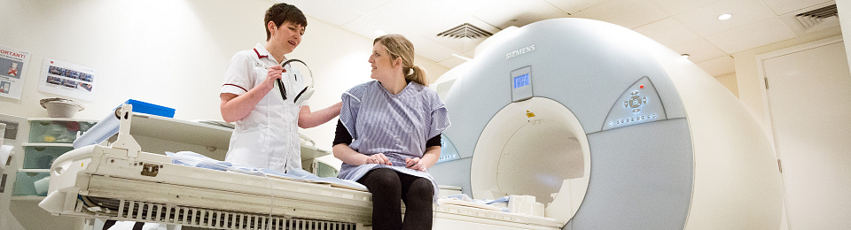
PET imaging is widely used in the management of cancer patients. Most commonly, an FDG PET scan is carried out to identify areas with high glucose metabolism, such as tumours. These images are useful for diagnosis, staging and monitoring treatment.
Such a scan requires the injection of a radioactive ‘tracer’ – which is taken up by the tumour tissue – and therefore the procedure has an associated radiation dose for the patient and for staff at the imaging facility.
A recent study by scientists at Central Manchester University Hospitals NHS Foundation Trust and The University of Manchester investigated whether technological developments in scanner equipment over the last decade could allow a reduction in the amount of radioactive tracer used.
Ian Armstrong, a nuclear medicine physicist who led the study, said: “Despite improvements in PET technology, we haven’t seen any change in the guidance regarding the amount of injected radiotracer we should use for FDG PET.”
PET imaging relies on the detection of simultaneous pairs of gamma rays produced when positron particles emitted by the injected tracer interact inside the body. The team looked at an analysis approach using time-of-flight (TOF) information, which utilises the faster detectors present in modern PET systems to more accurately locate the source of each pair of rays.
They found that by making use of TOF information, they could reduce the number of ‘counts’, or individual gamma ray pairs, they measured. This means that for the same quality of image, they could reduce the injected radioactive dose, or scan for a shorter period of time.
Ian said:
Here in Manchester we’ve decided to use this improvement to do both – reduce the administered activity and the scan time. As a result we have managed to lower the radiation dose for cancer patients and our staff and also increase the numbers of scans we are able to carry out.
Paper entitled “The assessment of time-of-flight on image quality and quantification with reduced administered activity and scan times in 18F-FDG PET” Armstrong et al. (2015) Nuclear Medicine Communications 36:728-37
Cancer is one of The University of Manchester’s research beacons – examples of pioneering discoveries, interdisciplinary collaboration and cross-sector partnerships that are tackling some of the biggest questions facing the planet.